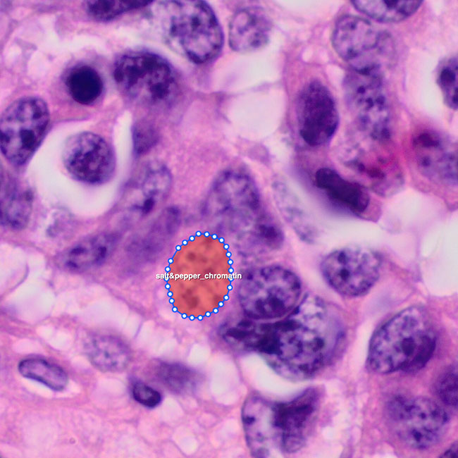
Prostate Cancer
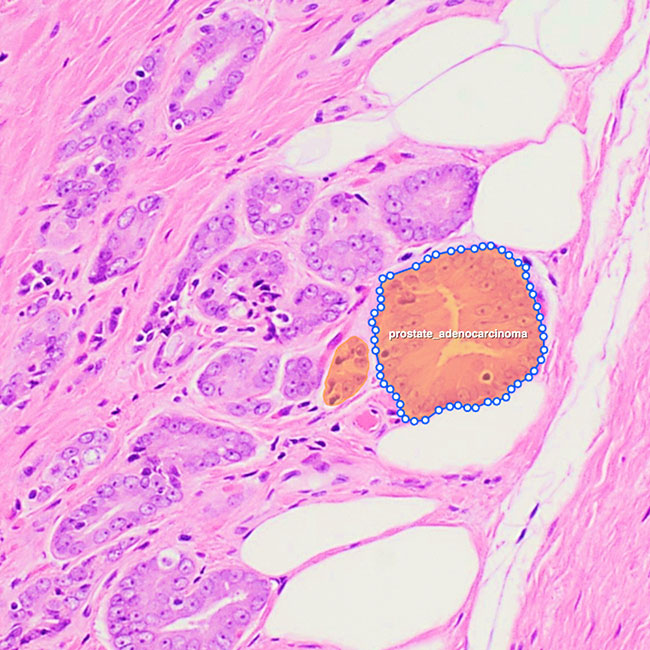
Annotators delineate glandular structures and tumor regions on prostate core biopsies and whole-slide images. Pathologist-led QA ensures clinically accurate Gleason grading and helps train AI models for early detection and grading.
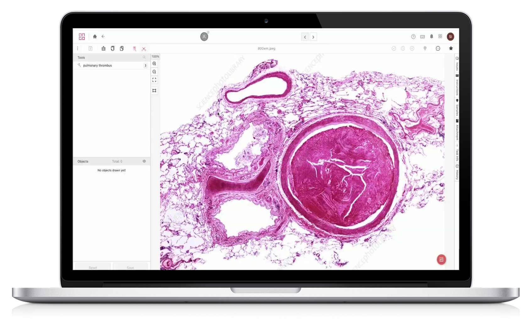
Whole Slide Image (WSI) Labeling with High Accuracy to Accelerate AI-Assisted Pathologic Diagnoses

AI and computer vision models can now easily differentiate between normal and abnormal tissues and even identify patterns too complex for the human eye in just a fraction of the time.
However, these models commonly rely on understanding tissue structural and cytologic variations in whole slides. The accuracy and reliability of results depend on cost-effective approaches to create high-volume and high-quality annotated pathology datasets.
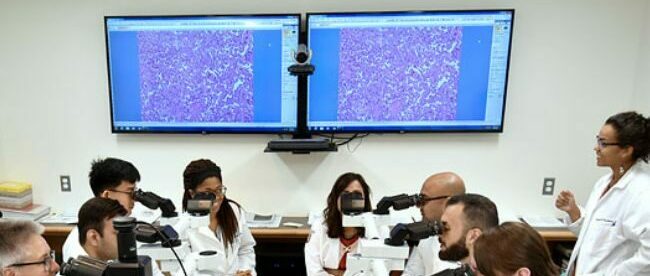
iMerit’s pathologist-led specialized teams annotate cells and tissues on whole slide images to help academic centers, pharmaceutical companies, and digital pathology startups develop next-generation diagnostic products and treatments across a host of diseases.
Our extensive tool ecosystem with custom Expert-in-the-loop workflows helps achieve cost-efficient scaling while being HIPAA-compliant and conforming to FDA regulatory approval standards.
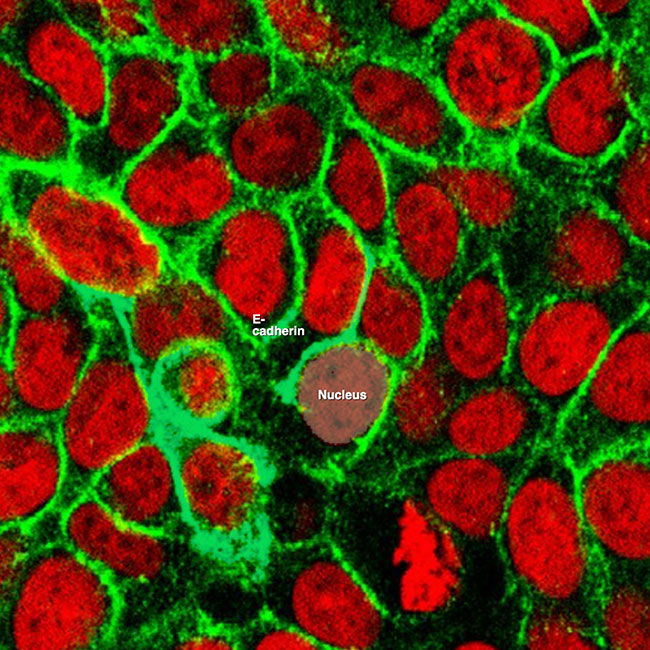
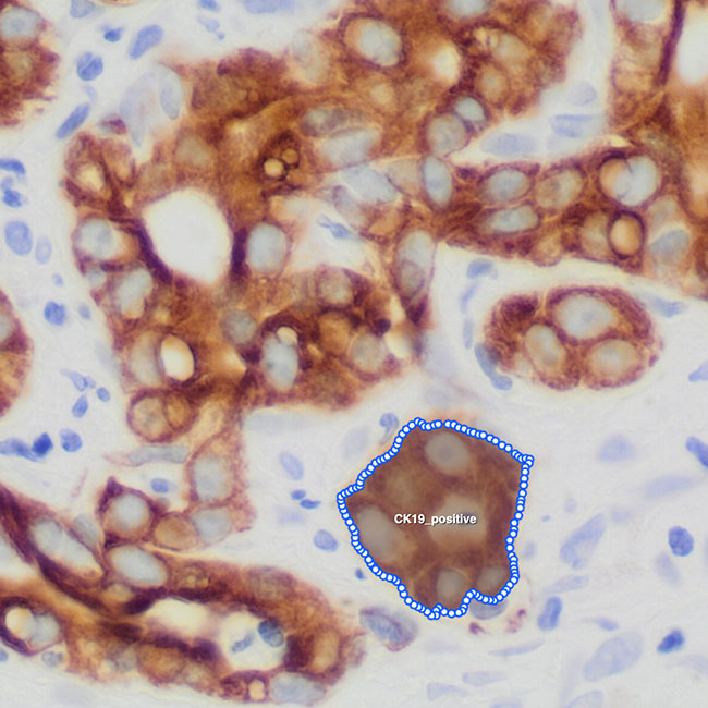
iMerit supports AI/ML teams in the healthcare industry by accurately annotating cells and tissues across various traditional stains, including Immunohistochemistry (IHC), Chromogenic in situ hybridization (CISH), and Immunofluorescence (IF, FISH).
Mn
Data Points Enriched for Healthcare AI
%
Pathologist Productivity Gain through Diagnostic Support
%
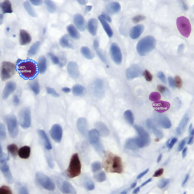
F1 Score vs. Expert Gold Set - Ki67 Breast Cancer Staining
A Major Public Health Institution Improves Cancer Treatment with iMerit
One of the challenges of this Public Health Institution, specializing in biomedical and health research, is that it took them almost two weeks to analyze biopsy reports, leading to increased patient emotional burden and a higher risk of disease progression.
With high-quality annotated images of cancer cells by iMerit’s team, the client could generate ground truth datasets for an AI model that added diagnostic support and triage to their clinicians.
Annotators delineate glandular structures and tumor regions on prostate core biopsies and whole-slide images. Pathologist-led QA ensures clinically accurate Gleason grading and helps train AI models for early detection and grading.
iMerit supports tumor detection and classification in lung biopsy slides. Our annotation teams segment tumor types (adenocarcinoma, squamous cell carcinoma) and support IHC-based biomarker quantification (PD-L1 expression and more).
Annotation teams classify epithelial tissue, detect invasive fronts, and segment polyps or adenocarcinoma regions. These datasets support AI tools for colorectal cancer screening and histological subtype identification.
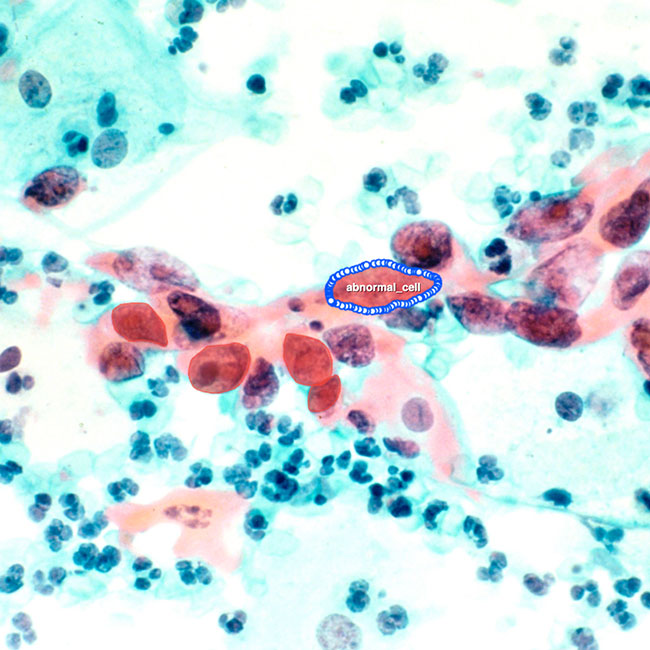
Using digital Pap smear slides, iMerit annotators label abnormal squamous and glandular cells. These annotations aid in training AI models for cervical dysplasia detection and HPV-related cancer risk stratification.
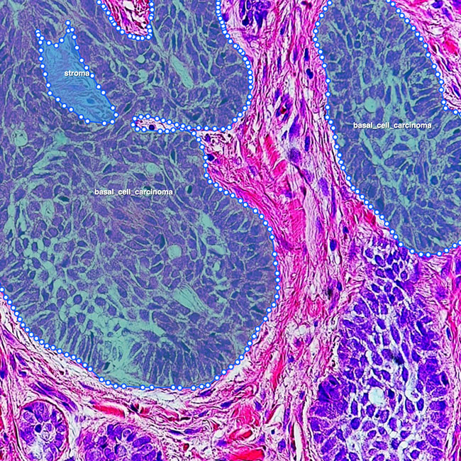
For dermatopathology use cases, iMerit supports annotation of melanocytic lesions, mitotic activity, and immune infiltration, helping build datasets for skin cancer detection and recurrence prediction.
Our teams annotate lymphoid structures and classify cell morphology to support lymphoma subtype classification. IHC biomarker quantification (CD20, Ki67 and more) assists in grading and monitoring progression.
iMerit annotates histopathological slides from liver biopsies to detect hepatocellular carcinoma and fibrosis staging. Annotations are used in AI development for early-stage HCC detection and treatment planning.
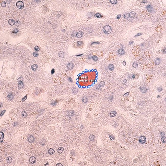
In addition to IHC staining, iMerit’s team supports detection of micrometastases in sentinel lymph nodes to assist in breast cancer staging and therapy decisions.
Our annotators support CNS tumor workflows, including glioma classification and tumor grading based on cell density, mitoses, and necrosis; often combining H&E with molecular markers like IDH1 or ATRX.
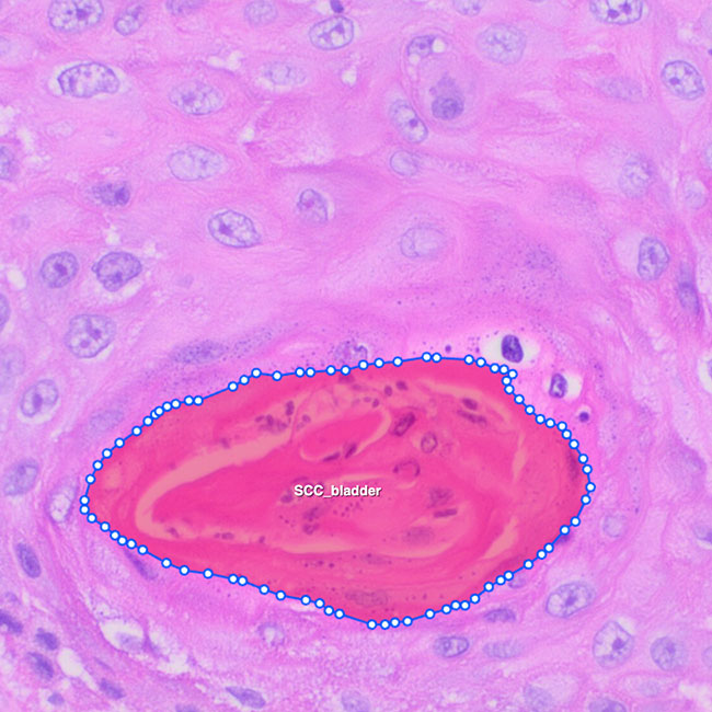
iMerit’s team provides tumor segmentation and grade classification for bladder carcinoma, often in transurethral resection specimens (TURBT), supporting AI models that guide therapeutic response.
"We have now done a more in-depth review of your work on the first phase of the annotation project, and we're delighted with the high level of accuracy."
Ph.D. Research Manager, Leading Medical Device Company