AI is helping close long-standing gaps in breast imaging, from mammography to MRI and ultrasound. But to move beyond research and into clinical practice, AI systems need more than high-level accuracy. They must be context-aware, workflow-ready, and clinically validated.
At iMerit, we support some of the most advanced diagnostic imaging AI teams by solving the real-world problems that arise when building scalable, regulatory-compliant tools. From complex DICOM workflows to multimodal annotation infrastructure, here is how we are helping shape the future of AI in breast imaging.
Comparing Time-Series Breast Images for Better Clinical Insights
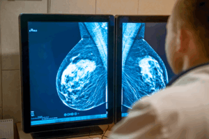
One of our recent clients needed to track changes in breast tissue over time by comparing a patient’s current and past mammograms side by side. This is critical for oncology professionals to assess treatment effectiveness and detect subtle disease progression.
To support this, our product and engineering teams upgraded the Ango Hub platform to allow simultaneous display of up to 16 DICOM images. This made it possible for radiologists and medical data labeling teams to perform time-based image comparisons without switching views or losing reference points. Expert annotators can now assess the evolution of findings more efficiently and with greater confidence.
To support this, our product and engineering teams upgraded the Ango Hub platform to allow simultaneous display of up to 16 DICOM images. This made it possible for radiologists and medical image annotation teams to perform time-based image comparisons without switching views or losing reference points. Expert annotators can now assess the evolution of findings more efficiently and with greater confidence.
Supporting Symmetry-Based Analysis in Mammography
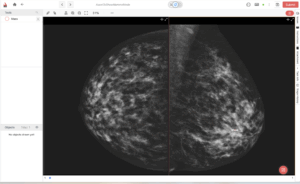
Breast symmetry is a key diagnostic factor in mammography. Our clinical partners highlighted the need for a tool that would let them compare left and right breast images with desired alignment to evaluate asymmetries more accurately.
In response, we developed a dedicated mammography mode within Ango Hub. This feature automatically snaps paired images to the midline, providing a mirrored, side-by-side view that makes it easier to detect structural differences and potential anomalies. This enhancement has been especially valuable in double-reading workflows and final QA reviews by radiology experts.
Measuring Angles in Breast Imaging Annotation
In breast imaging, subtle geometry can determine whether a lesion is flagged as suspicious or overlooked.
Why it matters for breast imaging AI:
- Symmetry assessment: Angle measurements enhance evaluation of asymmetries between left and right breasts, supporting early detection in line with MQSA and BI-RADS expectations.
- Lesion characterization: Radiologists can annotate the angular spread of calcification clusters or measure the spiculated extensions of a mass, directly supporting BI-RADS descriptors such as architectural distortion and spiculated margins.
- Implant and post-surgical cases: Measuring implant displacement angles or surgical changes provides structured data for complex scenarios that often challenge both radiologists and AI models.
- Regulatory-grade annotation: Numeric angle outputs create reproducible, standardized data that align with BI-RADS lexicon and strengthen audit trails for FDA and MQSA submissions.
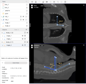
iMerit’s annotation workflows now include angle measurement tools that add quantitative precision to breast image labeling. By embedding quantitative geometry into breast imaging annotation, AI developers can train models that go beyond lesion detection to capture spatial characteristics and BI-RADS-aligned features, driving higher diagnostic accuracy and clinical trust.
Managing Complex Multimodal Inputs
Breast imaging AI is inherently multimodal. AI systems must process and annotate data from 2D mammograms, 3D tomosynthesis, breast ultrasound, and MRI, often in combination.
To handle this complexity, our teams use advanced toolchains that support multi-panel viewing, AI-assisted pre-labeling, and structured QA processes. We ensure that datasets remain consistent across modalities and that annotations follow clinical interpretation logic, not just visual cues. This is especially important for training models intended for regulatory submission.
Our Experts, Not the Crowd
None of this works without the right people. Our medical data labeling workforce includes MQSA-qualified radiologists, certified pathologists, and trained medical reviewers with experience in DICOM workflows and clinical documentation. We do not rely on crowdsourced labor. Every annotation project is guided by clinical standards and reviewed through expert-in-the-loop workflows that allow feedback and learning.
This quality-first approach is essential for AI systems moving toward FDA clearance or EU MDR compliance. It ensures that the datasets powering these systems reflect how real clinicians make decisions.
Beyond Labeling: Full-Lifecycle AI Support
Our involvement does not end at annotation. We support clients through model training, tuning, validation, and post-market updates. For teams building breast imaging AI, this means faster iteration cycles, more robust evaluations, and higher readiness for clinical integration.
The iMerit Advantage: What We’ve Learned
- Clinical context matters as much as data volume.
- Tooling must support the way radiologists actually work.
- Side-by-side comparison and symmetry analysis are not just convenience features; they are clinical requirements.
- Specialized workflows are needed to keep multimodal imaging aligned and consistent.
- Data quality improves dramatically when domain experts are involved from the start.
Driving the Future of Breast Imaging AI
From startups developing breast cancer detection algorithms to global platforms integrating multimodal diagnostic models, we are proud to support the teams making preventative care smarter and more accessible. Our mission is not just to provide labeled data. It is to help build the infrastructure that turns AI into a trusted partner in clinical care.
If you are building diagnostic models in breast imaging and facing challenges with data, tooling, or workflow design, we are here to help.
Explore iMerit’s Breast Imaging AI Solutions
Learn more about our diagnostic imaging expertise and advanced tooling here.
For platform capabilities, set up a demo or explore the Ango medical data labeling editor documentation to see how our tools support complex imaging tasks.
Want to discuss breast imaging annotation workflows? Contact us to see how we can help you build clinically precise, scalable AI solutions.