PathologyAnnotation
Whole Slide Image (WSI) Labeling with High Accuracy to Accelerate AI-Assisted Pathologic Diagnoses

Challenges of Pathology Annotation
AI and computer vision models can now easily differentiate between normal and abnormal tissues and even identify patterns too complex for the human eye in just a fraction of the time.
However, these models commonly rely on understanding tissue structural and cytologic variations in whole slides. The accuracy and reliability of results depend on cost-effective approaches to create high-volume and high-quality annotated pathology datasets.
iMerit Expertise
iMerit’s pathologist-led specialized teams annotate cells and tissues on whole slide images to help academic centers, pharmaceutical companies, and digital pathology startups develop next-generation diagnostic products and treatments across a host of diseases.
Our extensive tool ecosystem with custom Expert-in-the-loop workflows helps achieve cost-efficient scaling while being HIPAA-compliant and conforming to FDA regulatory approval standards.
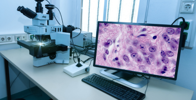
High-Quality Digital Pathology Annotation
iMerit supports AI/ML teams in the healthcare industry by accurately annotating cells and tissues across various traditional stains, including Immunohistochemistry (IHC), Chromogenic in situ hybridization (CISH), and Immunofluorescence (IF, FISH).
- iMerit’s workforce undergoes a rigorous pathologist-developed curriculum to deliver high-quality images efficiently.
- Specialized medical annotators work hand-in-hand with pathologists in hybrid workflows.
- US board-certified physicians are available for benchmarking and validation.
- HIPAA-compliant annotation processes will ensure data stays protected and secure.
- Our regulatory-grade processes ensure AI-assisted products get FDA 510K clearance.
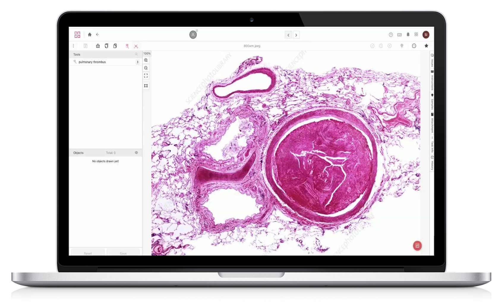
- iMerit’s tool set includes WSI functionality and Auto-labeling features.
20
Mn
Data Points Enriched for Healthcare AI
300
%
Pathologist Productivity Gain through Diagnostic Support
97
%
F1 Score vs. Expert Gold Set - Ki67 Breast Cancer Staining
Case Study

A Major Public Health Institution Improves Cancer Treatment with iMerit
One of the challenges of this Public Health Institution, specializing in biomedical and health research, is that it took them almost two weeks to analyze biopsy reports, leading to increased patient emotional burden and a higher risk of disease progression.
With high-quality annotated images of cancer cells by iMerit’s team, the client could generate ground truth datasets for an AI model that added diagnostic support and triage to their clinicians.
Common Use Cases
Leukemia
Using a 3-step workflow, iMerit annotators classify and segment cancer cells in bone marrow aspirates. The annotated images undergo a review by pathologists to ensure accuracy while being cost-effective.


Breast Cancer Immunohistochemistry
A consensus workflow to identify tumor boundaries help create a training dataset while minimizing biases and errors.

"We have now done a more in-depth review of your work on the first phase of the annotation project, and we're delighted with the high level of accuracy."
Ph.D. Research Manager, Leading Medical Device Company

Download Solution Brief